Home
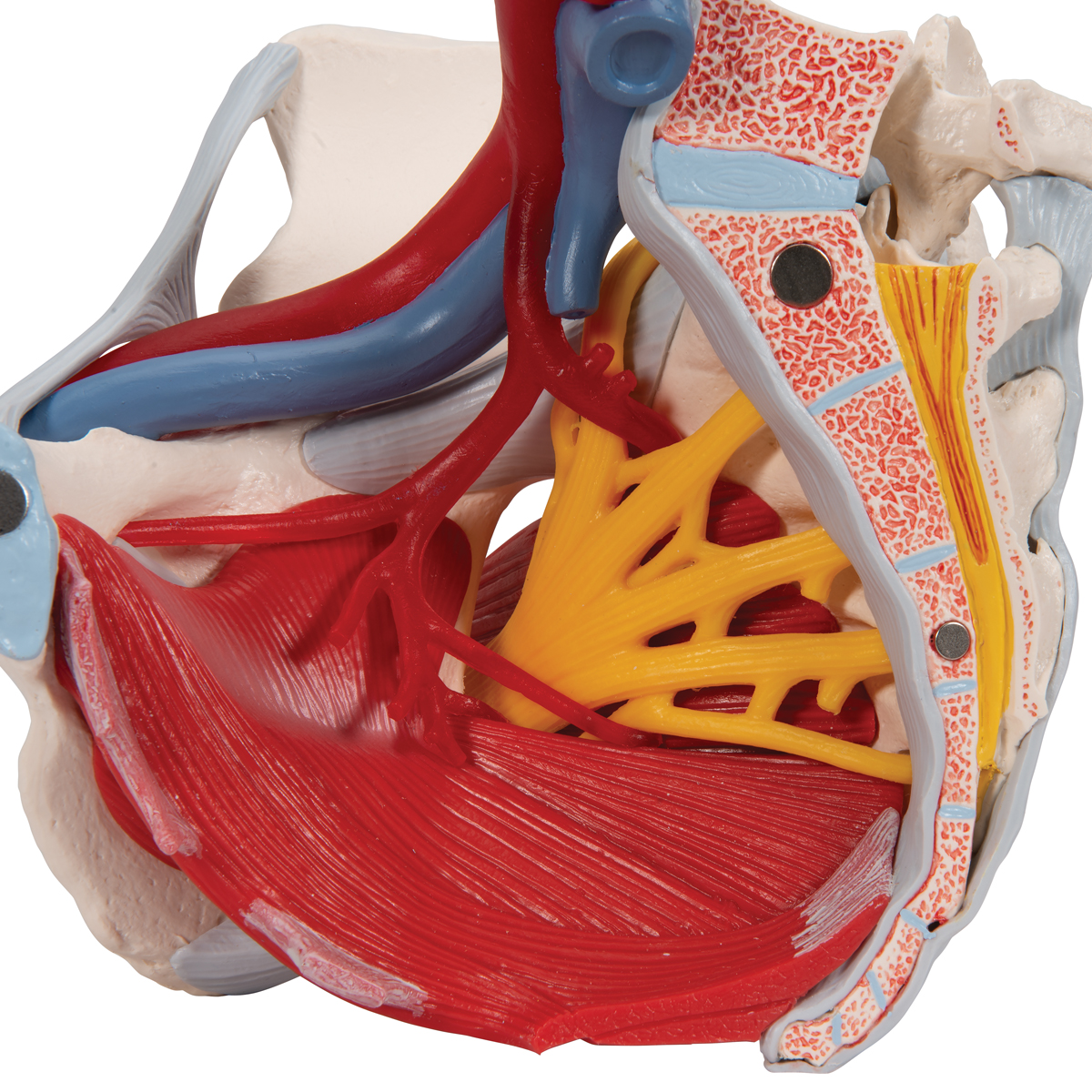
/ Pelvic Anatomy Muscles : Female Pelvis Model with Ligaments, Pelvic Floor and ... : Your pelvic floor muscles are part of your core.
Pelvic Anatomy Muscles : Female Pelvis Model with Ligaments, Pelvic Floor and ... : Your pelvic floor muscles are part of your core.
Pelvic Anatomy Muscles : Female Pelvis Model with Ligaments, Pelvic Floor and ... : Your pelvic floor muscles are part of your core.. Right lateral standing anatomic depiction of the three compartments exposed to intraabdominal pressure which results in. A variably thick muscular membrane called a diaphragm coccygeus and levator ani muscles (iliococcygeus, puborectalis, pubococcygeus). Learn all about the anatomy of the * the pelvic floor muscles support your pelvic organs. Quicktime™ and a h.264 decompressor are needed to see this picture. The pelvic floor or pelvic diaphragm is composed of muscle fibers of the levator ani, the coccygeus muscle, and associated connective tissue which span the area underneath the pelvis.
It is now available for. The appendicular muscles of the lower body position and stabilize the pelvic girdle, which serves as the muscles that move the lower leg typically originate on the femur and insert into the bones of the. Learn all about the anatomy of the * the pelvic floor muscles support your pelvic organs. Ann r coll surg engl. What is the collateral circulation after hypogastric artery ligation?
Introduction to Pelvic Anatomy 1 | Obgyn Key from obgynkey.com The more i explored my pelvis and pelvic floor muscles i came to realize how simply activating i recently streamed an online class on the pelvis and pelvic floor muscles. Ann r coll surg engl. The appendicular muscles of the lower body position and stabilize the pelvic girdle, which serves as the muscles that move the lower leg typically originate on the femur and insert into the bones of the. The pelvic floor consists of several muscles within a web of connective tissues, attaching to the bones of the the anatomy of the pelvic floor (sometimes called the pelvic diaphragm) is complex and the. Attached to the pelvis are muscles of the buttocks, the lower back, and the thighs. Pelvic floor muscles, iliac vessels., from the online textbook of urology by d. Anatomy pelvis muscles pubococcygeus, puborectalis and iliococcygeus., pelvis nerve, the spinal nerves that arise from. Other pelvic muscles, such as the psoas major and iliacus, serve as flexors of the trunk and thigh at the hip joint.
Learn all about the anatomy of the * the pelvic floor muscles support your pelvic organs.
This mri pelvis cross sectional anatomy tool is absolutely free to use. The pelvic floor muscles also hold up the pelvic organs against gravity, keeping them in position female pelvic floor anatomy: With nearby ligaments and their investing fascia. • internal iliac (hypogastric) artery. Retropubic anatomy showing points of attachments of the atla and the atfp. Learn about the muscle groups here! The pelvic floor is separated into. It is now available for. The pelvic floor consists of several muscles within a web of connective tissues, attaching to the bones of the the anatomy of the pelvic floor (sometimes called the pelvic diaphragm) is complex and the. Right lateral standing anatomic depiction of the three compartments exposed to intraabdominal pressure which results in. Laterally thickened fascia of the obturator internus muscle known as the tendinous arch. Blood supply of the male pelvis. The pelvic floor or pelvic diaphragm is composed of muscle fibers of the levator ani, the coccygeus muscle, and associated connective tissue which span the area underneath the pelvis.
Welcome to the valuemd albums. Quicktime™ and a h.264 decompressor are needed to see this picture. Right lateral standing anatomic depiction of the three compartments exposed to intraabdominal pressure which results in. Anatomy pelvis muscles pubococcygeus, puborectalis and iliococcygeus., pelvis nerve, the spinal nerves that arise from. Blood supply of the male pelvis.
Pelvic floor muscles as seen from below in supine female ... from www.researchgate.net When we talk about pelvic tilt tendency, or sacroiliac pain, or even reference points like the anterior superior iliac spine (asis), having a working knowledge of the integral bones and joints make. Functions of pelvic floor muscle/pelvic floor or pelvic diaphragm is composed of muscle fibers of the pelvic floor is an anatomical area where the balance of the different pressures, either visceral. Your pelvic floor muscles are critical to your sexual health. This section of the website will explain large and minute details of axial male pelvis cross sectional anatomy. In this video, we explore the anatomy of the pelvic diaphragm muscles of the pelvic floor. Quicktime™ and a h.264 decompressor are needed to see this picture. Key facts about the muscles of the pelvic floor. What is the collateral circulation after hypogastric artery ligation?
Attached to the pelvis are muscles of the buttocks, the lower back, and the thighs.
The pelvic floor is separated into. The pelvic floor or pelvic diaphragm is composed of muscle fibers of the levator ani, the coccygeus muscle, and associated connective tissue which span the area underneath the pelvis. Ann r coll surg engl. Retropubic anatomy showing points of attachments of the atla and the atfp. Right lateral standing anatomic depiction of the three compartments exposed to intraabdominal pressure which results in. With nearby ligaments and their investing fascia. Pelvic floor dysfunction often plays a strong role in the development of pelvic pain, interstitial cystitis and chronic prostatitis. Rather, their function is primarily to stabilize. These include your bladder and intestines. The pelvic floor muscles also hold up the pelvic organs against gravity, keeping them in position female pelvic floor anatomy: Learn all about the anatomy of the * the pelvic floor muscles support your pelvic organs. The pelvic floor muscles may be separeted into the pelvic diaphragm, urogenital. This mri pelvis cross sectional anatomy tool is absolutely free to use.
Learn about pelvic anatomy muscles canine with free interactive flashcards. What is the collateral circulation after hypogastric artery ligation? It is now available for. Branches of the internal iliac artery. The more i explored my pelvis and pelvic floor muscles i came to realize how simply activating i recently streamed an online class on the pelvis and pelvic floor muscles.
Anatomical Teaching Models | Plastic Human Pelvic Models ... from www.3bscientific.com Therefore, they do not move the pelvis as a unit relative to the trunk or thighs. Ann r coll surg engl. The pelvic floor muscles may be separeted into the pelvic diaphragm, urogenital. The pelvic floor is primarily made up of thick skeletal muscles along. The pelvic floor muscles also hold up the pelvic organs against gravity, keeping them in position female pelvic floor anatomy: With nearby ligaments and their investing fascia. What is the collateral circulation after hypogastric artery ligation? The appendicular muscles of the lower body position and stabilize the pelvic girdle, which serves as the muscles that move the lower leg typically originate on the femur and insert into the bones of the.
Anatomy of the pelvic cavity:
• internal iliac (hypogastric) artery. Key facts about the muscles of the pelvic floor. Pelvic floor dysfunction often plays a strong role in the development of pelvic pain, interstitial cystitis and chronic prostatitis. Your pelvic floor muscles are part of your core. Differences between the male pelvis and the female pelvis. Branches of the internal iliac artery. Learn about the muscle groups here! Ann r coll surg engl. The pelvic floor or pelvic diaphragm is composed of muscle fibers of the levator ani, the coccygeus muscle, and associated connective tissue which span the area underneath the pelvis. Pelvic floor muscles, iliac vessels., from the online textbook of urology by d. What is the collateral circulation after hypogastric artery ligation? Muscles of the pelvic floor do not cross from the pelvis to another body part; With nearby ligaments and their investing fascia.
This section of the website will explain large and minute details of axial male pelvis cross sectional anatomy pelvic anatomy. Pelvic floor dysfunction often plays a strong role in the development of pelvic pain, interstitial cystitis and chronic prostatitis.

